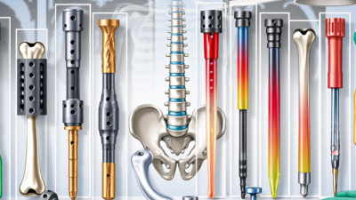
Top 10 Types of Maxillofacial Surgery Equipment Worldwide

Maxillofacial Surgery depends on precision, visibility, and controlled tissue handling. From trauma repair to corrective jaw procedures, surgeons work around bone, nerves, blood vessels, teeth, and delicate facial structures. The equipment must therefore support both clinical accuracy and patient safety.

Sir Harold Gillies, a pioneer in facial reconstruction, famously said, “The face is the most important part of the body.” His words still influence modern surgical planning. A millimeter can change a patient’s bite, breathing, speech, or appearance. That reality makes equipment selection more than a purchasing decision. It becomes part of treatment quality.

This guide examines the Top 10 Types of Maxillofacial Surgery Equipment Worldwide. It covers operating lights, surgical handpieces, bone saws, piezoelectric devices, surgical drills, retractors, fixation systems, navigation platforms, imaging equipment, and sterilization technology. Each category serves a specific purpose beside the operating table. A piezoelectric unit may reduce soft-tissue damage during bone cutting. A navigation system can display the instrument’s position near the orbit or skull base. A powerful light can reveal a narrow surgical field without creating distracting shadows.

Not every advanced device improves every procedure. That point deserves reflection. Equipment can be expensive, unfamiliar, or poorly matched to a hospital’s workflow. Experienced teams still need training, maintenance, and sound clinical judgment. The best technology is not always the newest model. It is the system that performs reliably, supports the surgical plan, and protects the patient throughout Maxillofacial Surgery.
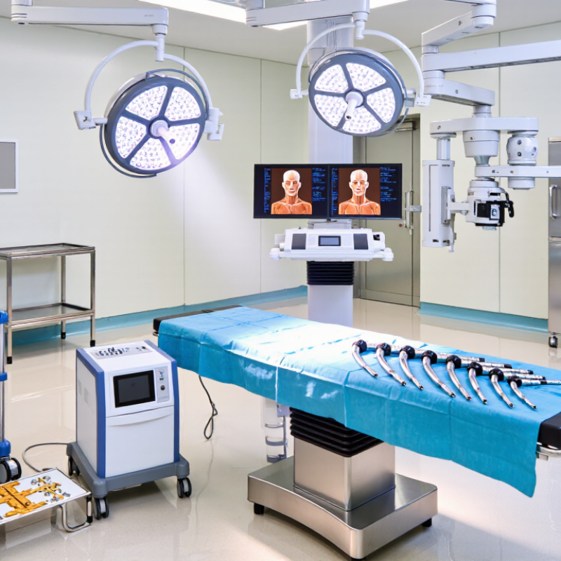
Top 10 Types of Maxillofacial Surgery Equipment Worldwide

Definition and Scope of Maxillofacial Surgery Equipment

Maxillofacial surgery equipment refers to the instruments, devices, and systems used to examine, plan, perform, and monitor facial surgery. It supports procedures involving the jaw, mouth, facial bones, soft tissues, salivary glands, and related structures. The scope begins before the incision. Imaging units, examination lights, surgical microscopes, and digital planning tools help clinicians understand anatomy and treatment risks. Some tools are simple. Others integrate software, sensors, and three-dimensional models.

During surgery, the category includes cutting and dissecting instruments, retractors, suction systems, powered handpieces, fixation tools, and electrosurgical devices. Anesthesia workstations, patient monitors, operating tables, and sterile storage also influence procedural safety. Reconstruction may require measuring guides, bone contouring tools, and systems that support precise placement of fixation components. These devices do not replace clinical judgment. Training, maintenance, sterilization, calibration, and emergency readiness remain essential. Small failures matter.

Postoperative scope includes recovery monitors, wound-care equipment, and imaging used to assess healing or complications. However, classification can vary between hospitals and regulatory systems. A fixation plate is an implant, not strictly equipment, although its placement depends on specialized instruments. This boundary deserves careful explanation in product research. Reliable evaluation considers accuracy, ergonomics, compatibility, cleaning requirements, operator training, local requirements, and documented clinical performance. Specifications alone can mislead. A device that cleans poorly may delay the next case.

Diagnostic and Imaging Equipment for Maxillofacial Procedures

Diagnostic and Imaging Equipment for Maxillofacial Procedures

Diagnostic imaging guides many maxillofacial procedures, from implant planning to facial trauma assessment. Cone-beam computed tomography provides three-dimensional views of bone, teeth, and nearby anatomical structures. It can reveal canal positions that ordinary images may hide. Panoramic systems offer a broad view of both jaws with relatively simple positioning. Cephalometric imaging supports facial growth analysis and orthognathic surgery planning. Intraoral scanners capture detailed digital impressions without traditional impression materials.

Accuracy depends on more than the equipment itself. The operator must position the patient carefully and select an appropriate exposure setting. Small head movements can blur important borders. Metal restorations may create streaks or dark areas. These artifacts can mislead inexperienced users. No scan is perfect. A useful image still requires clinical examination and professional judgment.

In a reliable workflow, qualified radiology professionals review the images and document their findings. Surgeons should compare scans with symptoms, physical findings, and previous records. DICOM compatibility supports secure image transfer and long-term case review. Regular calibration, maintenance logs, and staff training help preserve consistent quality. Yet protocols can become too rigid. A low-dose scan may be safer, but it might not answer every surgical question. The imaging choice should match the patient, the procedure, and the information genuinely needed.

Top 10 Types of Maxillofacial Surgery Equipment Worldwide - Diagnostic and Imaging Equipment for Maxillofacial Procedures
Rank Equipment Type Imaging or Detection Principle Common Maxillofacial Applications Typical Output Radiation Profile Main Clinical Strength Important Limitation or Consideration
1 Cone-Beam Computed Tomography (CBCT) System Cone-shaped X-ray beam with rotational acquisition and three-dimensional reconstruction. Dental implant planning, impacted teeth, orthognathic surgery, craniofacial trauma, jaw lesions, temporomandibular joint assessment, and airway evaluation. High-resolution multiplanar and 3D volumetric images. Ionizing X-rays Detailed bone visualization with generally lower radiation and smaller equipment requirements than conventional medical CT. Soft-tissue contrast is limited; dose varies substantially with field of view, exposure settings, and protocol selection.
2 Panoramic Dental X-Ray System Rotational two-dimensional X-ray projection of the jaws and dentition. Initial assessment of teeth, mandible, maxilla, eruption patterns, fractures, gross lesions, and preoperative dental screening. Single curved panoramic image of both jaws. Ionizing X-rays Broad coverage with a relatively quick examination and comparatively low dose. Magnification, distortion, superimposition, and limited depth information reduce precision for complex surgical planning.
3 Intraoral Digital Radiography System Small intraoral detector records X-ray projections of selected teeth and supporting structures. Periapical pathology, dental infections, periodontal bone levels, root morphology, localized trauma, and endodontic evaluation. High-detail two-dimensional intraoral radiographs. Ionizing X-rays Excellent fine-detail resolution for individual teeth and localized alveolar bone. Small field of view and two-dimensional superimposition limit evaluation of extensive facial anatomy.
4 Multidetector Medical Computed Tomography (MDCT) Scanner Multiple detector rows measure X-ray attenuation to create cross-sectional and volumetric images. Complex facial fractures, polytrauma, deep-space infection, skull-base disease, advanced tumors, and urgent postoperative assessment. Rapid multiplanar, 3D, and bone-window reconstructions; contrast-enhanced studies when indicated. Ionizing X-rays Fast, wide anatomical coverage and strong depiction of bone and acute traumatic abnormalities. Higher radiation exposure than many dental imaging examinations; iodinated contrast may be needed for selected soft-tissue studies.
5 Cephalometric X-Ray System Standardized lateral or posteroanterior radiographic projection of the skull and facial skeleton. Orthodontic and orthognathic planning, facial growth analysis, skeletal relationships, and preoperative cephalometric measurements. Standardized lateral or frontal skull radiograph, often with tracing software. Ionizing X-rays Reproducible linear and angular measurements for facial skeletal and dental relationships. Two-dimensional projection causes superimposition and does not provide complete three-dimensional anatomical detail.
6 Magnetic Resonance Imaging (MRI) System Magnetic fields and radiofrequency pulses generate images from tissue-specific proton signals. Temporomandibular joint discs, masticatory muscles, salivary glands, nerves, marrow, soft-tissue tumors, and postoperative soft-tissue assessment. Multiplanar high-contrast soft-tissue images; specialized sequences may include diffusion or angiographic information. No ionizing radiation Superior soft-tissue contrast and useful visualization of structures that are poorly shown on dental radiographs. Longer examination time, higher cost, motion sensitivity, noise, and safety restrictions for some implants or retained metal.
7 3D Facial Surface Scanner Optical structured light, stereophotogrammetry, or related camera-based surface capture. Facial asymmetry analysis, soft-tissue documentation, reconstructive planning, orthognathic simulation, and postoperative outcome comparison. Three-dimensional color or texture-mapped facial surface model. No ionizing radiation Non-contact recording of external facial form with useful surface measurements and visual comparison. Does not show teeth roots, bone, or deep soft tissues; accuracy can be affected by movement, hair, reflective surfaces, and facial expression.
8 Intraoral Optical Scanner Optical image capture and software stitching create a digital surface model of teeth and oral tissues. Digital impressions, restorative and prosthetic workflows, orthodontic records, implant planning, occlusion analysis, and surgical guide design. Color or monochrome 3D digital dental arch and occlusal model. No ionizing radiation Improves digital workflow efficiency and avoids conventional impression material for many applications. Captures visible surfaces rather than internal anatomy; scan accuracy may decline across long spans, saliva, blood, or limited access.
9 Maxillofacial Ultrasound System High-frequency sound waves are transmitted and received to display tissue interfaces and movement. Superficial facial masses, salivary glands, lymph nodes, vascular lesions, soft-tissue foreign bodies, and image-guided aspiration or biopsy. Real-time two-dimensional images, with optional Doppler flow information. No ionizing radiation Portable, dynamic, relatively low-cost imaging that can support real-time procedures. Bone and air block sound transmission, so deep facial spaces, teeth, and most intracranial structures are not well assessed.
10 Digital Maxillofacial Photography System Standardized visible-light image capture using controlled positioning, lighting, and scale. Clinical documentation, facial analysis, wound and scar monitoring, treatment communication, surgical planning records, and outcome assessment. Standardized extraoral and intraoral two-dimensional photographs. No ionizing radiation Inexpensive, accessible, and effective for documenting visible soft-tissue changes over time. Provides no direct information about bone, tooth roots, internal lesions, or three-dimensional depth without additional imaging.
Ranking reflects a practical overview of broad clinical usefulness, availability, and relevance to maxillofacial diagnosis and surgical planning; it is not a market-share ranking. The appropriate modality depends on the clinical question, patient factors, radiation justification, and local imaging protocols.

Cutting, Drilling, and Bone Shaping Systems

Top 10 Types of Maxillofacial Surgery Equipment Worldwide

Cutting, drilling, and bone shaping systems support precise work in maxillofacial surgery. Common categories include oscillating saws, reciprocating saws, rotary drills, surgical burs, micro-saws, piezoelectric cutters, powered rasps, bone shapers, ultrasonic tools, and irrigation-assisted handpieces. Each system suits different bone density, surgical access, and anatomical risk.

Cutting systems should provide stable control without excessive vibration. Oscillating and micro-saws can create controlled osteotomies, while piezoelectric devices may help protect nearby soft tissue.

Rotary drills and burs remove bone efficiently, but heat remains a serious concern. Continuous irrigation, sharp instruments, and measured pressure help reduce thermal injury. The operator must also monitor visibility and tissue movement.

Bone shaping systems refine edges after major cuts. Powered rasps and finishing burs can smooth irregular surfaces around reconstructive sites. Torque control matters during drilling, especially near thin cortical bone. Sterilization quality, handpiece balance, noise, and maintenance affect real-world performance. A detailed specification sheet cannot predict every operating-room challenge. Technique still matters more than speed.

There is room for error.

Even a precise device can remove too much bone when visibility is poor. Teams should match equipment settings with anatomy, imaging, and surgical training. A cautious pace often produces a more reliable contour.

Soft-Tissue, Microsurgical, and Implant Placement Instruments

Maxillofacial surgery equipment must match tissue behavior, surgical access, and placement accuracy.

Soft-tissue instruments include fine scissors, tissue forceps, needle holders, and retractors. Their narrow profiles reduce crushing and improve visibility around delicate mucosa. A surgeon may prefer a slightly curved instrument when working near the oral commissure or nasal lining.

Small details matter.

A balanced handle can reduce wrist strain during long procedures. Microsurgical instruments demand finer control. Micro-forceps, needle holders, dilators, and bipolar coagulation tools support vessel repair and nerve-related procedures.

Their tips must align precisely, without excessive pressure. Even a minor bend can affect suturing accuracy. That mistake is easy to overlook.

Implant placement instruments form another major category.

Osteotomy drills, depth gauges, parallel pins, torque-controlled drivers, and insertion tools help guide implants into planned positions. Surgical guides may improve consistency, but they cannot replace anatomical judgment.

Bone density, limited opening, and unexpected defects can change the procedure. The equipment should therefore be selected alongside imaging quality, sterilization capacity, and staff training.

Reusable instruments require documented inspection, cleaning, packaging, and sterilization cycles. A clean-looking instrument may still carry hidden damage or residue.

Global teams also need to verify local regulatory requirements and validated maintenance procedures.

No instrument guarantees a predictable result. Careful handling, continuous training, and honest review of complications remain essential.

Navigation, Safety, and Digital Technologies in Maxillofacial Surgery

Maxillofacial surgery equipment now connects navigation, imaging, safety, and digital planning. Optical and electromagnetic navigation systems can guide osteotomies near the orbit, skull base, and mandibular canal. Cone-beam CT adds three-dimensional detail while reducing dependence on two-dimensional assumptions. The screen is not the patient. Registration accuracy still depends on stable reference points, clear anatomy, and careful verification.

The World Health Organization reports that one in ten patients experiences harm in healthcare, with more than half considered preventable (WHO Patient Safety Fact File, 2023). This finding gives surgical safety equipment a practical purpose. Intraoperative imaging, instrument tracking, airway monitoring, and digital checklists can reduce avoidable uncertainty. However, technology can also create false confidence. A loose tracker, outdated scan, or poorly calibrated device may mislead an experienced team.

Digital workflows extend beyond navigation. Three-dimensional planning, patient-specific guides, surgical simulation, and additive manufacturing support more predictable reconstruction. Market research reports continue to show strong growth in surgical navigation and medical imaging technologies, reflecting wider hospital investment in precision care. Yet implementation remains uneven. Training, maintenance, cybersecurity, and data governance require equal attention. That gap matters. The best equipment cannot replace anatomical judgment, direct visual inspection, and disciplined communication.