What Is Arthroscopic Surgery and How Does It Work?
Arthroscopic Surgery is a minimally invasive technique used to examine and treat problems inside a joint. Instead of opening the joint widely, a surgeon makes small incisions near the affected area. A slender camera, called an arthroscope, enters through one incision. Its lighted lens sends magnified images to a monitor in the operating room.
Orthopedic surgeon Dr. James R. Andrews describes arthroscopy as a way to “visualize, diagnose, and treat problems inside a joint.” That idea explains the procedure’s practical value. The surgeon may identify damaged cartilage, a torn ligament, inflamed tissue, or loose fragments. Small instruments can then repair or remove the problem through additional openings. The knee and shoulder are common treatment areas. The ankle, hip, wrist, and elbow may also be treated.
The process can look simple on a screen.
It is not simple surgery.
Before recommending Arthroscopic Surgery, a specialist reviews symptoms, physical findings, scans, activity levels, and medical history. Sometimes an arthroscope confirms what imaging only suggested. In other cases, it reveals less damage than expected. That uncertainty matters. A smaller incision does not guarantee a faster recovery or a perfect result. Swelling, stiffness, infection, nerve irritation, and continued pain remain possible. Recovery also depends on rehabilitation and the underlying condition.
This guide will explain how arthroscopy works, why doctors use it, and what patients may experience before, during, and after treatment. It will also examine its limits, because informed decisions require more than attractive surgical images.
What Arthroscopic Surgery Is and Why It Is Performed
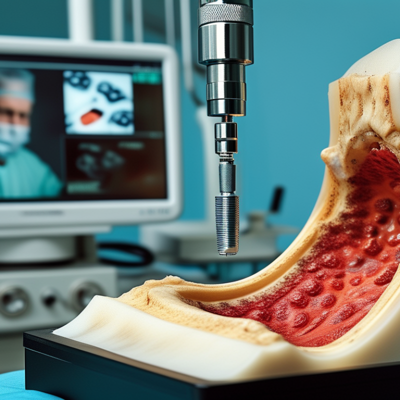
Arthroscopic surgery is a minimally invasive procedure used to examine and treat problems inside a joint. A surgeon inserts a small camera, called an arthroscope, through a tiny incision near the joint. The camera sends magnified images to a monitor, showing cartilage, ligaments, tendons, and synovial tissue. Additional small openings may hold slender instruments for treatment. This approach usually causes less muscle disruption than open surgery, but it is still real surgery.
Doctors may recommend arthroscopy when pain, swelling, stiffness, or limited movement persists after examination and non-surgical care. It can help diagnose and treat certain meniscus tears, ligament injuries, loose cartilage, inflamed tissue, and some shoulder conditions. The purpose depends on the joint, imaging results, symptoms, and daily activities. A scan alone does not always explain the pain. That matters.
During the operation, anesthesia prevents pain, while sterile fluid gently expands the joint for clearer viewing. The surgeon may repair tissue, remove loose fragments, or smooth a damaged surface. Recovery varies widely. Some patients walk soon after knee arthroscopy, while others need crutches, physical therapy, and gradual strengthening. Possible risks include infection, bleeding, blood clots, nerve irritation, stiffness, and continuing symptoms. Arthroscopy is not automatically better for every condition. I would not treat a promising scan as a guarantee of relief.
How the Arthroscopic Procedure Is Prepared and Performed
Arthroscopic surgery uses a small camera and narrow instruments through tiny skin openings. The surgeon views the joint on a monitor instead of making a large incision. It commonly treats damaged cartilage, torn ligaments, inflamed tissue, and loose fragments.
Preparation begins with a careful medical review. Patients should disclose allergies, blood-thinning medicines, diabetes, sleep apnea, and previous anesthesia problems. The team confirms the correct joint, reviews scans, explains expected benefits, and discusses alternatives. Fasting instructions depend on the anesthesia plan. A nurse also checks skin condition and infection risks. The World Health Organization reported that surgical-site infections affect about 2–5% of surgical patients in high-income settings. Clean skin and strict sterile technique matter.
On the operating table, anesthesia keeps the patient comfortable. The surgeon cleans the joint area and creates portals, often only a few millimeters wide. Sterile fluid gently expands the joint, improving visibility. The camera reveals the internal surfaces. Instruments may remove damaged tissue, smooth cartilage, repair a tear, or reconstruct a structure. A WHO surgical-safety evaluation found major complications fell from 11% to 7% after checklist use. Small steps can carry serious weight.
Afterward, staff monitor breathing, pain, swelling, and movement. Crutches or a brace may be needed. Recovery depends on the procedure, not merely the incision size. That distinction is easy to miss. Arthroscopy is less invasive, but it is not risk-free. Persistent fever, worsening redness, severe calf pain, or unusual drainage requires prompt medical assessment. Different hospitals organize preparation differently, and that variation deserves honest discussion.
What Happens Inside the Joint During Arthroscopy
Arthroscopic surgery uses a narrow camera to examine and treat a joint through small skin openings. The procedure commonly involves the knee, shoulder, hip, wrist, or ankle. Anaesthesia reduces pain while the patient remains carefully monitored.
Inside the joint, the surgeon gently inserts the arthroscope through a small incision. Sterile fluid flows into the space, expanding it and improving visibility. The camera sends magnified images to a monitor.
On screen, cartilage looks smooth and pale, while inflamed tissue may appear swollen or reddened. The surgeon can inspect ligaments, tendons, cartilage, and the joint lining.
Small instruments enter through separate openings. They may remove loose cartilage, trim damaged tissue, repair a ligament, or smooth rough surfaces. Every movement is controlled and deliberate. The joint is rinsed before the instruments are removed, and the incisions usually need only simple closure.
The screen shows important details, but it cannot explain every source of pain. A clear image is not a complete answer. Some damage may look minor yet cause significant symptoms, while visible changes may cause none.
Careful examination, medical history, and imaging still matter. Swelling, stiffness, infection, bleeding, or nerve irritation can occur after surgery, although serious complications are uncommon. Recovery depends on the treated joint, the procedure, and consistent rehabilitation. Progress is rarely perfectly straight.
Common Uses and Conditions Treated with Arthroscopy
What Is Arthroscopic Surgery and How Does It Work?
Common Uses and Conditions Treated with Arthroscopy
Arthroscopy is a minimally invasive procedure used to examine and treat a joint. A surgeon inserts a small camera through a tiny incision. The camera displays enlarged images on a monitor. Additional instruments enter through nearby openings. This approach may reduce tissue damage compared with traditional open surgery.
Doctors commonly use arthroscopy for persistent knee pain, torn menisci, and damaged cartilage. It can also treat anterior cruciate ligament injuries, though reconstruction plans vary. In the shoulder, arthroscopy may repair rotator cuff tears, labral injuries, and recurrent dislocations. Other applications include ankle cartilage damage, hip impingement, and elbow problems.
Not every joint complaint needs surgery. A careful examination and imaging review should guide the decision. Symptoms can come from arthritis, inflammation, or referred pain. Arthroscopy may not relieve pain caused mainly by advanced arthritis. That distinction is sometimes underestimated.
During recovery, patients may use crutches, a sling, or supervised exercises. Recovery time depends on the joint and the procedure performed. A simple inspection may heal quickly. Ligament repair usually requires several months of rehabilitation. Swelling, stiffness, infection, blood clots, and nerve irritation remain possible risks. Results are not guaranteed. A second clinical opinion can clarify uncertain cases.
Recovery, Benefits, Risks, and Possible Limitations
Arthroscopic surgery uses a small camera to view a joint through tiny incisions.
The surgeon inserts narrow instruments through nearby openings. A monitor guides each movement inside the joint. This approach may treat damaged cartilage, torn ligaments, or inflamed tissue. It is not always suitable. Recovery often begins at home, sometimes within hours. Patients may use crutches, ice packs, and prescribed pain relief. Swelling can make stairs uncomfortable during the first few days. Physical therapy usually restores strength, movement, and confidence. Healing times differ widely.
Smaller incisions may reduce tissue damage and shorten recovery. They can also leave smaller scars. However, less visible damage does not guarantee less pain. Possible risks include infection, bleeding, stiffness, and blood clots. Nerve or blood vessel injury is uncommon but possible. Anesthesia can cause temporary nausea or dizziness. Serious complications are rare, but no operation is risk-free. A patient’s age, general health, joint condition, and therapy effort affect results.
Arthroscopy cannot reverse advanced arthritis or repair every type of joint damage.
Some pain may remain after technically successful surgery. Imaging and physical examination help determine whether surgery is appropriate. A careful discussion should cover expected benefits, recovery milestones, and alternatives. Ask what success means in your specific case. Results can be less predictable than expected. That uncertainty deserves honest attention.