What Is Fracture Fixation and What Types Are Available?
Fracture Fixation is the surgical stabilization of a broken bone while it heals. It may use plates and screws along the bone, a metal nail inside its canal, or pins connected to an external frame. Small wires can also hold selected fractures in place. The choice depends on the fracture’s location and pattern, the surrounding soft tissue, and the patient’s health and daily needs. A fracture near a joint, for example, may need careful alignment so the joint surface heals smoothly.
AO pioneer Martin Allgöwer helped advance a principle often summarized as: “Fracture treatment should restore function, not just bone alignment.” That idea matters. A clear X-ray is useful, but it is not the whole story. Surgeons also consider stability, blood supply, pain, and how safely a patient can begin moving again. Not every fracture needs an operation, and no fixation method suits every person. The best plan can be less obvious than a textbook diagram suggests.
This guide introduces common fixation types and explains how they differ. It also looks at what may influence a surgeon’s recommendation, what recovery can involve, and which questions patients may want to ask. A plate under the skin, a nail inside the bone, or an external frame can each solve different problems. The details matter. So does honest discussion of risks, limits, and alternatives with a qualified orthopaedic professional.
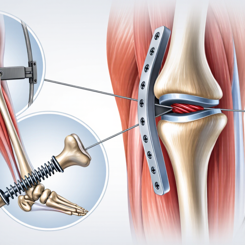
How Fracture Fixation Stabilizes Broken Bones
What Is Fracture Fixation and What Types Are Available?
How Fracture Fixation Stabilizes Broken Bones
Fracture fixation holds broken bone pieces steady and in useful alignment while healing begins. A 2021 Global Burden of Disease analysis published in The Lancet Healthy Longevity estimated 178 million new fractures worldwide in 2019. That scale helps explain why reliable stabilization matters. Surgeons may use screws and plates along a bone, a metal rod inside a long bone, or pins connected to an external frame. The choice depends on the fracture’s location, shape, and surrounding soft tissue.
Fixation limits unwanted movement, reduces the chance that fragments shift, and can help people begin appropriate movement sooner. Some screws compress a fracture; plates may support several pieces like a small bridge. But hardware does not heal bone by itself. Blood supply, bone quality, and the patient’s overall health matter too. An X-ray may show good alignment, yet recovery can still be uneven. That part deserves honest attention.
Tips: Follow your care team’s weight-bearing instructions. Ask when to check the incision and which symptoms need prompt review. Don’t adjust activity based only on pain; healing takes time.
When Fracture Fixation Is Recommended
Fracture fixation may be recommended when broken bone ends are displaced, unstable, or unlikely to stay aligned in a cast or brace. A surgeon may also consider it when a fracture crosses a joint and could affect smooth movement, or when several broken pieces make alignment difficult. The aim is to restore position and provide enough stability for healing. It depends.
The decision often draws on X-rays or CT scans, the injury mechanism, and the person’s health and daily needs. For example, a fracture that shifts when a patient moves may need fixation, while a stable fracture may heal well without surgery. Open fractures require urgent medical care; fixation may be part of treatment after the wound and surrounding tissue are assessed. Not every serious-looking image means an operation is best.
There is no tidy rule that fits every fracture. The choice can depend on swelling, skin condition, bone quality, and whether the person can safely follow movement restrictions. A clinician should explain expected benefits, risks, and alternatives, including possible infection, stiffness, or further procedures. Sometimes the plan changes after swelling settles or new imaging clarifies the pattern. That uncertainty can be frustrating, but it is worth discussing before treatment begins.
Principles That Guide Fracture Fixation
Fracture fixation stabilizes broken bone fragments so they can heal in a useful position. The method depends on the fracture pattern, bone quality, soft-tissue condition, and the patient’s overall health. A plate and screws may support a fracture near a joint, while an intramedullary nail can stabilize some long-bone breaks. Pins or an external frame may suit selected cases. These are options, not a ranking.
Good fixation aims to restore alignment and provide enough stability for healing without unnecessarily limiting nearby joints. The surgeon also considers blood supply, muscle and skin condition, and how much weight the bone can safely bear during recovery. A simple-looking break may still need careful planning. And plans can change: swelling, imaging findings, or unexpected bone quality may affect the approach. The trade-offs are not always neat.
Tips: Ask what movement and weight-bearing are safe, and when follow-up imaging is planned. Keep the wound clean as instructed, and report increasing redness, drainage, fever, or worsening pain promptly. Healing takes time. Do not adjust activity based only on how comfortable the limb feels.
Types of Internal and External Fixation
Fracture fixation keeps broken bone ends aligned while healing begins. Internal fixation places devices beneath the skin, directly on or inside the bone. A surgeon may use a metal plate with screws for a fracture near a joint, or a long intramedullary nail inside the hollow center of a thigh or shin bone. Small wires or pins can hold selected fragments, especially in hand and wrist injuries. The choice depends on fracture shape, bone quality, nearby joints, and soft-tissue damage. Details matter.
External fixation uses pins or wires that pass through the skin and connect to a frame outside the body. The frame can stabilize a badly injured limb while allowing access to wounds and swollen tissue. It may be temporary, with internal fixation considered later, or remain in place until the bone heals. Pin sites need careful cleaning and monitoring for redness, increasing pain, or drainage. They can feel awkward, and everyday movement may need adjustment.
No method fits every fracture. Internal devices can irritate nearby tissue or require another procedure, while external frames may limit comfort and carry pin-site infection risks. A surgeon considers imaging, circulation, skin condition, and the patient’s overall health before recommending fixation. Even then, plans can change as swelling settles or the fracture’s stability becomes clearer. Recovery is not always tidy.
What Is Fracture Fixation? Internal and External Types
Fracture fixation stabilizes broken bone fragments while they heal. Internal fixation uses implants placed in or on the body, such as plates and screws or intramedullary nails. External fixation uses pins or wires connected to a frame outside the body. In this chart, 1 means a method belongs to that category; 0 means it does not. The values classify methods and do not represent frequency or treatment outcomes.
How Surgeons Choose a Fixation Method
Fracture fixation is not a hardware contest. Surgeons match the method to the fracture’s location, shape, stability, and surrounding tissue. A CT scan may reveal a split hidden on X-rays; a swollen, bruised limb can also affect timing. Bone quality, age, activity, other illnesses, and expected ability to bear weight matter. Plates and screws can control fragments directly, while an intramedullary nail sits inside a long bone. External fixation may provide temporary stability when soft tissue is badly injured. No scan decides alone.
Evidence helps, but it does not erase individual differences. In the 2017 Lancet FAITH trial, 1,079 adults with femoral-neck fractures received either a sliding hip screw or cancellous screws. Reoperation occurred in about 20% and 22%, respectively; the difference was not statistically significant. That result applies to this specific fracture group, not every broken bone. Surgeons also weigh alignment, blood supply, healing risk, surgical access, and how much stability is needed for safe movement. Sometimes the choice is less certain than patients expect.
Tips: Ask what the fixation is meant to achieve, when weight-bearing can begin, and what signs require follow-up. Share medicines and medical conditions honestly; small details can change the plan.