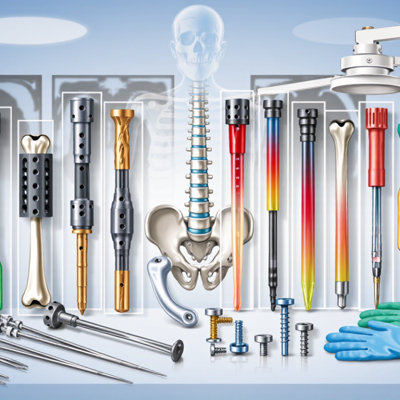
Choosing the right fracture fixation system can influence alignment, stability, healing, and a patient’s return to daily activity. Yet there is no universal “best” implant for every fracture. Bone quality, fracture pattern, soft-tissue damage, infection risk, age, and surgical experience all affect the decision. This guide examines What Are the 10 Best Fracture Fixation Systems? through practical and evidence-informed criteria.
The discussion includes locking plates, intramedullary nails, external fixators, screws, wires, and hybrid constructs. Each system has a specific role. A locking plate may support fragile metaphyseal bone, while an intramedullary nail can share load inside a long bone. External fixation may provide valuable temporary stability when swelling or open wounds limit internal surgery. Small details matter, including screw trajectory, implant fit, fluoroscopic views, and postoperative weight-bearing plans. They can change outcomes.
Look closely at the evidence.
Reliable selection requires more than comparing product names. It involves peer-reviewed research, regulatory clearance, manufacturer instructions, surgeon training, and long-term follow-up. Complications such as malalignment, nonunion, hardware irritation, and infection deserve honest attention. Some newer designs appear impressive but have limited independent data. That uncertainty matters. The “best” system may be the one that matches the injury and can be applied safely by an experienced team. Patients should discuss imaging findings, expected recovery, alternatives, and possible revision surgery with a qualified orthopedic specialist. This overview supports informed conversations, but it cannot replace individualized clinical judgment.
The 10 best fracture fixation systems cannot be ranked without defining the fracture first. AO/OTA classification provides that shared language. It records the bone, anatomical segment, fracture pattern, and complexity. A simple extra-articular fracture demands a different strategy from an unstable multifragmentary injury. Classification is a map, not a verdict. Radiographs can mislead. Computed tomography may reveal joint extension, hidden fragments, or rotational instability.
Six criteria should guide fixation selection: fracture morphology, soft-tissue condition, bone quality, required stability, patient factors, and surgical resources. Fracture morphology influences whether a plate, nail, screw construct, wire, or external frame fits the mechanical problem. Soft-tissue swelling may favor limited exposure or temporary stabilization. Poor bone quality requires stronger purchase and careful load sharing. Stability needs vary between compression, bridging, and controlled motion. Patient age, activity, healing risks, and comorbidities also matter. Finally, the system must match available imaging, instruments, training, and follow-up.
In practice, the “best” construct is often the least complicated one that restores alignment and protects biology. That sounds obvious. It is not always easy. Surgeons may overvalue familiar hardware or attractive technical features. I have also seen classification used too rigidly, ignoring patient-specific anatomy. A reliable decision records the AO/OTA pattern, compares fixation options, and explains why one method offers safer control with fewer compromises.
What Are the 10 Best Fracture Fixation Systems?
Map the 10 Systems: Plates, Nails, Screws, Wires, and External Frames
The 2019 Global Burden of Disease Study estimated 178 million new fractures worldwide. Published in The Lancet Public Health, it linked fractures with rising disability and healthcare demand. That burden makes fixation selection a clinical question, not a popularity contest. This map covers ten systems across plates, nails, screws, wires, and external frames.
Compression plates bring fragments together, while locking plates preserve alignment in weaker bone. Intramedullary nails sit inside the canal and share load along the limb. They can help with many shaft fractures, but anatomy changes the decision. Screw systems include cortical, cancellous, cannulated, and headless compression designs. Cortical screws purchase dense bone. Cancellous screws use deeper threads in softer metaphyseal bone. Cannulated screws guide placement over a wire. Headless screws can reduce surface prominence.
K-wires offer simple control for small fragments or temporary stabilization. Tension-band wiring converts muscle pull into compression. Its performance may weaken with migration, irritation, or poor tensioning. External frames stabilize complex injuries while keeping hardware away from damaged skin. The 2023 MarketsandMarkets report estimated the orthopedic trauma devices market at about 8.8 billion dollars. It projected growth to roughly 12.4 billion dollars by 2028. That forecast describes commercial demand, not better patient outcomes. No ranking survives every fracture pattern. Surgeons may combine plates and screws, wires and frames, or revise plans during surgery. That variability is easy to understate.
A practical map of plates, nails, screws, wires, and external frames by their primary mechanical role.
The relative scores summarize established mechanical characteristics: load sharing, resistance to bending, control of compression or tension, and suitability for temporary or definitive stabilization. They are educational comparisons rather than clinical rankings; implant selection depends on fracture pattern, bone quality, soft-tissue condition, and surgeon judgment.
Choosing among the ten best fracture fixation systems requires more than comparing catalog features. The fracture pattern, bone quality, soft-tissue condition, and patient activity all change the decision. In practice, I would compare five biomechanical measures before calling any system “best.”
Construct stiffness shows how much the fixation resists bending under everyday forces. Interfragmentary motion indicates whether movement may support healing or threaten stability. Axial load capacity measures resistance to compression, especially during weight bearing.
Torsional strength reveals how well the construct handles twisting. Fatigue strength matters because implants experience repeated loading, not one dramatic event. Screw pullout resistance also deserves attention, although it is influenced by bone density and placement technique.
These measures overlap, but they are not interchangeable. A rigid construct is not automatically the safest choice.
Tips: Review radiographs in multiple views, then match stability to the fracture’s biological needs. Check working length, screw distribution, fixation points, and expected loading. Laboratory values help, but they cannot fully predict clinical healing. Small details matter. I have seen elegant plans weaken when bone quality was underestimated. That is the part worth questioning. Final selection should follow current clinical evidence, surgical training, patient-specific assessment, and careful follow-up.
The ten best fracture fixation systems cannot be chosen by hardware strength alone. Clinical value appears in four outcomes: union, reoperation, infection, and mechanical failure. A useful review compares similar fractures, follow-up periods, and patient risk profiles. A tibial shaft fracture in a healthy adult differs greatly from an open, contaminated injury. That distinction matters. Published studies may report union above 90%, yet definitions and imaging schedules often vary.
Intramedullary nails commonly support early loading and may reduce soft-tissue disruption. Locking plate constructs can provide stable fixation near joints, but wound complications remain important.
External fixation can protect damaged tissues, although pin-site infection and later procedures may affect results. Hybrid and fragment-specific systems may improve control in complex patterns, but evidence is often based on small series.
I would hesitate to call one system universally best. Surgeon experience, reduction quality, bone health, smoking, diabetes, and injury severity can change every rate.
Tips:
Check how each study defines union. Separate superficial from deep infection. Record planned and unplanned reoperations. Examine failure timing, not only failure totals. Look for at least one year of follow-up. Be cautious when studies exclude high-risk patients. A low failure rate may reflect careful selection, not superior hardware. Clinical judgment still has imperfect edges.
What are the 10 best fracture fixation systems? The answer changes with anatomy, bone quality, and soft-tissue damage. For the wrist, a volar locking plate suits many displaced distal-radius fractures. A dorsal spanning plate can protect highly comminuted injuries, but wrist stiffness remains a concern. The CDC reports that over 3 million older adults receive emergency care for fall injuries annually. That makes low-profile, reliable wrist fixation especially important.
The forearm often benefits from a compression plate, while selected fractures may accept an intramedullary forearm nail. For the humerus, a locking plate offers direct control, and an antegrade humeral nail limits exposure. At the ankle, an anatomically contoured locking plate can support unstable malleolar fragments. A separate syndesmotic screw construct may restore distal tibiofibular stability. These choices require careful imaging, not habit.
For the tibia, an interlocking tibial nail is useful for many shaft fractures and allows earlier movement. A circular external fixator remains valuable when swelling or open soft-tissue injury limits internal fixation. Pelvic injuries may need a reconstruction plate for accessible anterior-ring fractures, or a percutaneous iliosacral screw system for selected posterior injuries. The WHO Global Status Report on Road Safety 2023 estimates 1.19 million annual road-traffic deaths worldwide, reinforcing the need for adaptable trauma systems. My practical bias favors the least invasive stable option, although that judgment can fail when CT findings, bone density, or surgeon experience are underestimated.
| Fracture Fixation System | Typical Strengths and Best Use | Wrist | Forearm / Elbow | Shoulder / Proximal Humerus | Ankle | Hip / Proximal Femur | Pelvis / Acetabulum |
|---|---|---|---|---|---|---|---|
| Volar Locking Plate | Fixed-angle support for articular or metaphyseal fractures, especially in osteoporotic bone. | Preferred Common for unstable distal radius fractures, including many intra-articular patterns. |
Selective May be used for selected distal forearm fractures; not a routine choice for shaft fractures. |
Limited Not designed for proximal humerus fixation. |
Limited Not a standard ankle construct. |
Limited Not appropriate for proximal femoral load transfer. |
Limited Not intended for pelvic or acetabular fixation. |
| Dorsal Spanning Plate | Temporary bridge fixation that maintains length and alignment across a severely comminuted wrist. | Preferred Useful for complex distal radius fractures, fracture-dislocations, and injuries unsuitable for conventional plating. |
Limited Generally not used for forearm or elbow fractures. |
Limited Not a shoulder fixation system. |
Limited Not intended for ankle stabilization. |
Limited Not used for hip or proximal femur fractures. |
Limited Not suitable for pelvic fixation. |
| Intramedullary Nail | Load-sharing fixation that preserves soft-tissue blood supply and supports long-bone alignment. | Limited Selected wrist applications exist, but plating is more common for distal radius fractures. |
Preferred Common for diaphyseal fractures of the radius, ulna, or humerus when anatomy and implant design are suitable. |
Selective Used in selected proximal humerus or humeral shaft fractures. |
Preferred Useful for selected distal tibia or fibula fractures, depending on fracture level and soft-tissue condition. |
Preferred Used for subtrochanteric and many diaphyseal femur fractures; design determines proximal femur suitability. |
Limited Not a primary system for most pelvic ring or acetabular fractures. |
| Locking Compression Plate | Versatile plate fixation offering compression, bridging, or fixed-angle locking for metaphyseal and shaft fractures. | Preferred Useful for selected distal radius, distal ulna, and complex wrist fractures. |
Preferred Common for forearm shaft fractures and selected distal humerus or periarticular injuries. |
Preferred Frequently used for proximal humerus and clavicle-region fracture patterns when plating is indicated. |
Preferred Common for distal fibula, distal tibia, and selected malleolar fractures. |
Selective Used for selected proximal femur, shaft, and distal femur fractures; implant contour and biomechanics are critical. |
Preferred Useful for selected anterior pelvic ring, iliac, and acetabular fractures with adequate exposure. |
| Monolateral External Fixator | Rapid, minimally invasive stabilization when swelling, contamination, or physiologic instability limits internal fixation. | Preferred Useful for highly swollen, open, or unstable distal radius fractures, often as temporary or definitive treatment. |
Selective Used for open fractures, severe soft-tissue injury, or staged treatment around the elbow and forearm. |
Selective May stabilize proximal humerus or humeral shaft injuries when internal fixation is unsafe. |
Preferred Common for temporary stabilization of severe open ankle or pilon fractures. |
Selective Can provide damage-control stabilization for femoral or hip-region injuries in critically ill patients. |
Preferred Important for temporary anterior pelvic stabilization in unstable pelvic ring injuries. |
| Circular Ring External Fixator | Multiplanar fixation with adjustable alignment and strong support for complex, infected, or limb-reconstruction cases. | Limited Rarely selected because less invasive wrist-specific options are usually available. |
Selective Useful for complex forearm reconstruction, bone loss, malunion, or infection. |
Limited Uncommon for isolated shoulder fractures. |
Preferred Useful for complex pilon fractures, bone loss, infection, and deformity correction. |
Selective May be used for complex femoral reconstruction, nonunion, or infection rather than routine hip fractures. |
Limited Not a routine definitive system for most pelvic ring or acetabular fractures. |
| Tension-Band / Cerclage Construct | Converts tensile forces into compression across selected simple fractures or supports fragments around an implant. | Selective May support selected ulnar styloid or small avulsion fragments, but is not a primary distal radius system. |
Preferred Common for simple olecranon fractures when the extensor mechanism and fracture geometry are suitable. |
Selective May be used for selected greater tuberosity or pat-like traction fragments, but not most proximal humerus fractures. |
Selective Can be used for selected medial or lateral malleolar avulsion patterns. |
Preferred Cerclage may supplement fixation around the proximal femur; tension-band principles apply to selected trochanteric fragments. |
Selective May assist fixation of selected iliac or pubic fragments, but is rarely sufficient alone. |
| Cannulated Screw Fixation | Accurate interfragmentary compression for simple fractures, osteochondral fragments, and selected periarticular injuries. | Selective Used for selected scaphoid, radial styloid, or simple intra-articular fragments. |
Selective Useful for selected radial head, coronoid, or simple periarticular fracture fragments. |
Selective May fix selected greater tuberosity or humeral head fragments when adequate purchase is possible. |
Preferred Common for medial malleolar, posterior malleolar, talar, and selected syndesmotic-related fracture components. |
Preferred Common for femoral neck fractures in younger or physiologically active patients with suitable fracture geometry. |
Preferred Useful for selected sacroiliac, trans-sacral, pubic ramus, and acetabular fragment fixation. |
| Sliding Hip Screw | Controlled collapse and compression for stable or selected unstable extracapsular proximal femur fractures. | Limited Not applicable to wrist fractures. |
Limited Not applicable to forearm or elbow fractures. |
Limited Not applicable to shoulder fractures. |
Limited Not applicable to ankle fractures. |
Preferred Established option for many stable intertrochanteric fractures; less suitable for subtrochanteric extension or some unstable patterns. |
Limited Not intended for primary pelvic ring or acetabular fixation. |
| Cephalomedullary Nail | Intramedullary load-sharing fixation for proximal femur fractures, especially patterns with instability or subtrochanteric extension. | Limited Not applicable to wrist fractures. |
Limited Not applicable to forearm or elbow fractures. |
Limited Not applicable to shoulder fractures. |
Limited Not applicable to ankle fractures. |
Preferred Common for unstable intertrochanteric, reverse-obliquity, and subtrochanteric femur fractures. |
Limited Not a primary pelvic or acetabular fixation system. |
| Pelvic and Acetabular Screw–Plate Construct | Contoured fixation and percutaneous or open stabilization of pelvic ring and acetabular fracture patterns. | Limited Not intended for wrist fractures. |
Limited Not intended for forearm or elbow fractures. |
Limited Not intended for shoulder fractures. |
Limited Not intended for ankle fractures. |
Limited Not intended for primary hip or proximal femur fixation. |
Preferred Used for selected anterior ring, posterior ring, iliac, and acetabular fractures based on stability and displacement. |