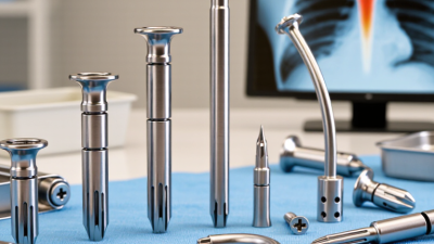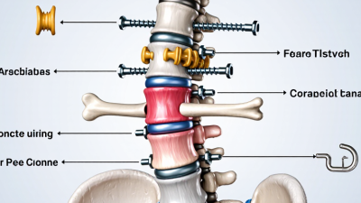
Intramedullary Nails are orthopedic implants placed inside the central canal of a broken bone. Surgeons commonly use them to stabilize femur, tibia, and humerus fractures. The nail follows the bone’s natural axis, while locking screws control rotation and length. This design can share weight across the fracture and support earlier movement when healing permits.
Real cases are messier than diagrams. A high-energy accident may leave crushed tissue, swelling, or several fracture lines. An orthopedic surgeon reviews X-rays and, when needed, computed tomography scans before selecting the nail’s size, entry point, and locking pattern. Treatment also depends on bone quality, infection risk, soft-tissue condition, and the patient’s general health. Intramedullary Nails may also support selected nonunions or certain pathological fractures, but their use requires careful diagnosis and specialist judgment.
The procedure is often less disruptive than exposing the entire fracture, yet it is not automatically simple or risk-free. Pain, infection, blood clots, malalignment, delayed healing, and hardware irritation can occur. Even experienced teams must adapt when anatomy or fracture behavior differs from expectations. That limitation matters. A nail can stabilize a fracture, but biology still has to repair it. Clinical evidence supports intramedullary fixation for many long-bone injuries, while guidelines and local practice may vary. Patients should discuss imaging findings, rehabilitation milestones, weight-bearing limits, and warning signs with their treating clinician. This article explains what these implants do, why surgeons choose them, and where their benefits may not apply.
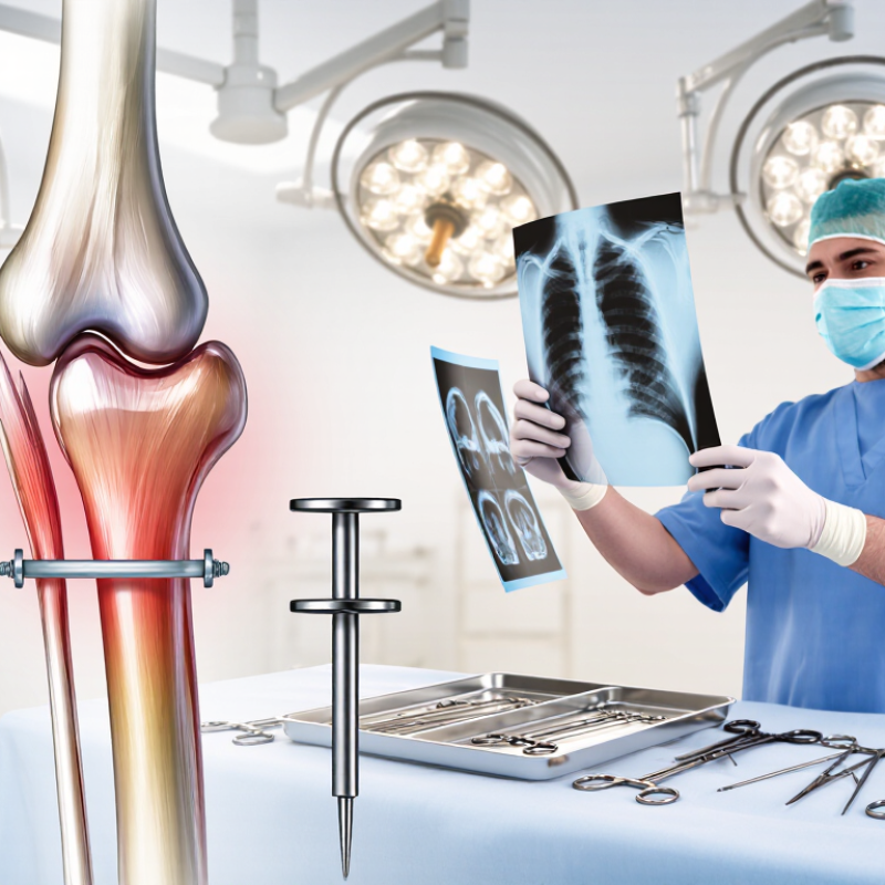
An intramedullary nail is a metal rod placed inside the hollow center of a broken bone. This inner space is called the medullary canal. Surgeons commonly use these implants for fractures of the thighbone, shinbone, and upper arm bone. The nail supports the bone from within, rather than sitting only along its outer surface.
During surgery, the medical team guides the nail through the bone canal and checks its position with live imaging. Locking screws near both ends can prevent unwanted rotation or shortening. This design often allows controlled weight sharing while the fracture heals. The exact nail size, entry point, and screw arrangement depend on the injury, bone shape, and the patient’s overall health.
The concept sounds simple. Healing is not. An intramedullary nail does not repair damaged tissue by itself. It mainly provides stability while the body builds new bone. Surgeons also consider blood supply, soft-tissue damage, infection risk, and alignment before choosing this method. Recovery may involve pain management, wound checks, physical therapy, and gradual weight-bearing. Some fractures heal slowly, and plans may change after follow-up images. That uncertainty deserves honest discussion. Pain, swelling, or unusual drainage should be reported promptly, even when the incision appears small.
| Data Dimension | Key Information | Typical Use and Practical Considerations |
|---|---|---|
| Definition | An intramedullary nail is a rigid implant placed inside the medullary canal of a long bone. | It acts as an internal splint, helping maintain alignment and stability while the fracture heals. The nail is commonly secured with locking screws at one or both ends. |
| Primary Purpose | Fracture fixation and restoration of bone alignment, length, and rotation. | Intramedullary nailing is used when a fracture requires surgical stabilization or when nonoperative treatment is unlikely to provide adequate alignment or functional recovery. |
| Femoral Shaft Fractures | Fractures through the central portion of the femoral shaft. | Antegrade or retrograde nailing may be selected according to the fracture location, associated injuries, and the surgeon’s approach. The implant provides strong mechanical support for a major weight-bearing bone. |
| Tibial Shaft Fractures | Fractures involving the shaft of the tibia. | Intramedullary nails are widely used for many displaced or unstable tibial shaft fractures. Careful assessment of soft-tissue injury and alignment is important because the tibia has limited soft-tissue coverage in several areas. |
| Humeral Shaft Fractures | Fractures through the shaft of the upper-arm bone. | Nailing can be considered for selected unstable, segmental, or multiple-trauma fractures. The treatment plan must account for shoulder function, elbow function, and nearby nerves. |
| Proximal or Distal Fractures | Fractures extending toward an end of a long bone. | Specialized nail designs and locking options may help treat fractures near the hip, knee, ankle, or shoulder. Fractures that extend into a joint may require another fixation strategy, depending on their pattern. |
| Open Fractures | Fractures associated with a wound that communicates with the fracture site. | Nailing may be used in selected cases after urgent wound evaluation, irrigation, debridement, antibiotic treatment, and assessment of the patient’s overall condition. Severe contamination or soft-tissue damage may change the timing or method of fixation. |
| Pathological Fractures | Fractures occurring in bone weakened by disease, such as certain tumors or metabolic conditions. | An intramedullary nail can provide load-sharing support and may help stabilize a bone at risk of fracture. Treatment also requires evaluation and management of the underlying disease. |
| Delayed Union or Nonunion | A fracture that heals more slowly than expected or fails to unite. | Exchange nailing, nail dynamization, bone grafting, or additional biological and mechanical treatments may be considered after the cause of poor healing has been evaluated. |
| Implant Design | Typically a hollow or solid metal rod with options for different lengths, diameters, curvature, and locking configurations. | The selected dimensions should match the patient’s anatomy and fracture pattern. Locking screws help control shortening, rotation, and axial movement. |
| Fixation Method | The nail is inserted through an entry point and may be locked proximally, distally, or at both ends. | Locked fixation is especially useful for unstable, comminuted, segmental, or length-unstable fractures. The entry point and surgical approach depend on the bone and nail design. |
| Biomechanical Advantages | The implant is positioned near the mechanical axis of the bone and can provide load-sharing stabilization. | Compared with some plates, intramedullary nails may reduce the need for extensive exposure of the fracture site and can support early movement when clinically appropriate. |
| Potential Benefits | Stable fixation, preservation of fracture-site blood supply in many cases, and support for earlier rehabilitation. | Possible benefits depend on fracture type, soft-tissue condition, bone quality, surgical technique, and the patient’s general health. Early weight-bearing is determined by the treating surgeon. |
| Potential Risks | Infection, malalignment, delayed healing, nonunion, hardware failure, blood loss, and injury to nearby nerves or vessels. | Other risks may include pain around the entry site, knee or shoulder symptoms, fat embolism, blood clots, and the need for revision surgery. Risk varies by injury and patient factors. |
| Postoperative Monitoring | Clinical examination and follow-up imaging are used to assess alignment, implant position, and fracture healing. | Patients are commonly monitored for wound problems, infection, neurovascular changes, increasing pain, loss of alignment, and signs of delayed healing. |
| Removal of the Nail | Routine removal is not required in every patient. | Removal may be considered for infection, prominent or symptomatic hardware, irritation, implant breakage, or other specific clinical reasons. The decision requires a risk–benefit assessment. |
| When It May Not Be Suitable | Certain fracture patterns, severe bone loss, active infection, unsuitable anatomy, or damage involving a joint may limit its use. | Alternative treatments may include plates and screws, external fixation, joint replacement, or staged reconstruction. The best method is selected after imaging and full clinical evaluation. |